| |
| |
|
Retinal
Detachment
This is a serious condition that if left untreated will
result in permanent blindness possibly with chronic
pain.
|
 |
|
How does it occur?
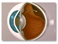
Retinal detachment is usually caused by the onset of
a tear or hole in the retina. Fluid then passes through
this hole from the vitreous gel into the space underneath
the retina lifting the retina off the wall of the eye
and resulting in loss of vision in the area that is
detached.
If this is treated in the early stages, vision can be
completely restored to normal, however if the central
vision is lost at the time of presentation to the doctor
then a variable amount of vision will be lost, depending
on the length of time and the degree to which the retina
is detached.
The results of surgery are generally very good -between
80 to 90% of retinas can be repaired with 1-2 operations.
Occasionally, particularly in the case where the detachment
is of long-standing nature or significant scar tissue
on the surface of the retina has formed, the prognosis
is not nearly as good.
The treatment includes one or more of the following
surgical options:
1) Laser treatment - can be done successfully
if the detachment is localized.
2) Pneumatic retinopexy - this involves the injection
of gas into the eye, the gas bubble within the eye will
then push the retina flat -this may require prolonged
positioning in different positions depending on where
the detachment is located -positioning may be upright,
on the side, or even face down. Positioning may last
between two to 10 days.
Approximately 60% of retinal detachments can be treated
in this manner.
3) Scleral buckling - this involves the placement
of a silicone band around the eye in order to indent
the wall of the eye. Intraoperative hemorrhage and other
complications including worsening of myopia or shortsightedness,
chronic ache around the eye, as well as occasional double
vision have been associated with scleral buckling.
4) Pars plana vitrectomy - involves the removal
of vitreous gel from the center of the eye. The tear
resulting the detachment is treated with laser after
removing fluid from beneath the retinal detachment.
Possible complications of this procedure include cataract
formation, infection-although the incidence of this
is very low - and failure of the procedure to achieve
fattening of the retina, which of course is a possibility
with any of the above maneuvers.
Peter
Hopp M.D.
|
|
|
| |
| |
|
|
|
|
|
Interior Retina, Kamloops, B.C.,
Canada, Dr. Peter Hopp, argon laser treatments for diabetic retinopathy,
branch retinal vein occlusions, clinically significant macular edema,
central serous retinopathy, lattice degeneration, macular edema
and retinal tears, retinal detachments, vitreous hemorrhages, dropped
nucleuses, macular holes
|
laser treatment of the retina,
laser treatment for glaucoma, laser treatment for diabetic eye disease,
laser treatment for certain types of macular degeneration,surgery
for cataracts, retinal detachment, macular hole, epiretinal membrane,
diabetic retinal disease,vitreous hemorrhages, chalazion excision,
entropion, other miscellaneous retinal and vitreous disorders
|
Interior Retina, Kamloops, B.C.,
Canada, Dr. Peter Hopp, argon laser treatments for diabetic retinopathy,
branch retinal vein occlusions, clinically significant macular edema,
central serous retinopathy, lattice degeneration, macular edema
and retinal tears, retinal detachments, vitreous hemorrhages, dropped
nucleuses, macular holes
|
|
Interior Retina provides treatment
and management of glaucoma, iritis, scleritis, vein/artery occlusions,
diabetic eye diseases, corneal abrasions, double vision, floaters,
optic neuritis, uveitis and after-cataracts.
|
|
|
|

